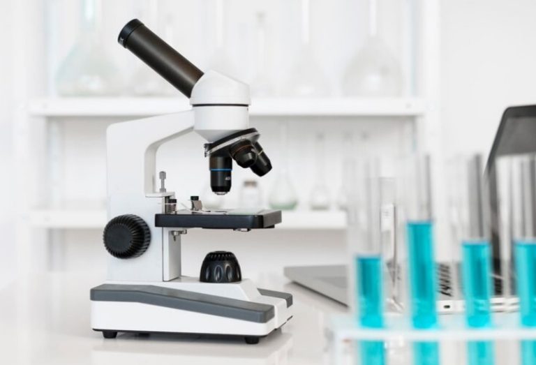
Microscopes are invaluable tools that enable scientists, researchers, and students to explore the microscopic world with precision and clarity. These devices have revolutionized our understanding of the intricate structures and processes that exist beyond the limits of our naked eye. At the core of any microscope are its essential components, which work in harmony to provide magnified views of specimens with remarkable detail. In this context, we will explore the three most important parts of a microscope: the objective lens, the eyepiece (ocular lens), and the illumination system. Understanding the functions and interplay of these components is crucial for harnessing the full potential of microscopy and unlocking the mysteries hidden within the minuscule realms of life and matter.
Three Most Important Parts of a Microscope
The three most important parts of a microscope are:
- Objective Lens: The objective lens is responsible for gathering light from the specimen and forming the primary image. It is usually located closest to the specimen and provides the highest magnification. Microscopes typically have multiple objective lenses with different magnification powers (e.g., 4x, 10x, 40x, 100x) that can be rotated into position depending on the desired level of magnification.
- Eyepiece (Ocular Lens): The eyepiece is the lens through which the viewer looks to observe the magnified image. It further magnifies the image produced by the objective lens. Eyepieces commonly have a magnification power of 10x, although other magnifications are also available. Microscopes may have a single eyepiece or binocular eyepieces for more comfortable viewing.
- Illumination System: The illumination system provides the necessary light to illuminate the specimen. It consists of a light source, condenser lens, and diaphragm. The light source can be a built-in bulb or an external source such as a halogen lamp or LED. The condenser lens collects and focuses the light onto the specimen, improving image clarity and resolution. The diaphragm controls the amount of light passing through the condenser, allowing adjustment of the brightness and contrast of the image.
While these three parts are essential for basic microscopy, it’s important to note that microscopes can have additional components such as a stage for holding the specimen, focus knobs for precise focusing, mechanical stages for easier specimen movement, and various adjustments to optimize image quality.
Conclusion
The objective lens, eyepiece (ocular lens), and illumination system stand as the foundational pillars of a microscope, collectively facilitating the visualization and exploration of the microscopic world. The objective lens captures light from the specimen, forming the primary image with varying levels of magnification. The eyepiece further magnifies this image, allowing the observer to perceive intricate details and structures. Meanwhile, the illumination system ensures adequate lighting, enhancing image clarity and contrast.
Together, these three components work in harmony to empower scientists, researchers, and students with the ability to unravel the hidden mysteries of cells, microorganisms, and tiny structures. With a firm grasp of these crucial parts, one can unlock the full potential of microscopy, fostering breakthroughs in various fields such as biology, medicine, materials science, and beyond. The continued advancement of these fundamental components, along with the integration of innovative technologies, will undoubtedly propel the field of microscopy to new horizons, expanding our understanding of the microscopic world and shaping the future of scientific discovery.